 Dr. Raymond Ribitch and his team use the latest in modern dental technology in their Mount Pleasant, MI office to ensure that they can accurately diagnose and treat dental problems. This technology also helps patients see and better understand the diagnostic process.
Digital Dentistry in Mid Michigan
Learn more about the dental technology we use below:
T-Scan
This diagnostic device records how a patient’s teeth touch and is used to detect bite issues. The T-Scan uses a sensor that the patient bites down on, and a computer analyzes the data, showing it in color in 2D or 3D graphics.
If only a few teeth touch, those teeth take the full force of a bite, which can result in damage. By detecting bite issues, our professionals are able to create a treatment that adjusts teeth for a balanced bite.
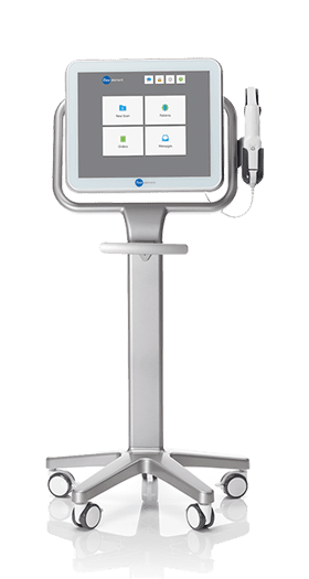
iTero
Gone are the days of messy dental impressions. The iTero scanner uses digital impressions that are wirelessly transmitted to a computer. The scanner captures detailed images of each tooth. These impressions are then used to create dental restorations, oral appliances, and Invisalign braces.
Intraoral Cameras
These small, lightweight cameras are used to show high-quality images of the mouth during treatment. These cameras are educational tools that allow patients a visualization of their procedure.
3-D Printing
Dr. Ribitch can create cost-effective temporary dental bridges and models of tooth crowns using a 3-D printer. 3-D printing is used for treatment planning in restorative and cosmetic treatments. To begin the 3-D printing process, Dr. Ribitch first takes digital scans of the mouth using an intraoral scanner. With these digital impressions, he can design a model of the teeth and prosthetics on a computer. After the design is complete, Dr. Ribitch sends it to the 3-D printer. The printer creates the model using high-quality resin. Once the model is printed, Dr. Ribitch cleans and prepares the resin.
Digital X-Rays
Digital x-rays, or radiographs, are used to find hidden dental structures, malignant or benign tumors, tooth decay, periodontal disease, and instances of bone loss.
Panoramic X-Rays
The panoramic radiograph creates a digital image of a patient’s upper and lower rows of teeth and jaws. It is used to detect sources of dental pain, temporomandibular joint abnormalities, areas of bone loss.
Cephalometric X-Rays
Cephalometric X-rays provide a radiograph of the side view of a patient’s head jaw and skull. This type of x-ray is most used for orthodontic treatments.
Spectra Laser Caries Detection
This device can help Dr. Ribitch identify dental caries or cavities in the mouth. The detection aid is non-invasive, using color and numerical indication to diagnose tooth decay.
If you suspect that you may have cavities, impacted teeth, or another dental problem, call Dr. Ribitch and his team at 989-772-1344.